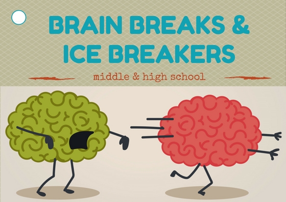
What is brain break
Brain Breaks Help Students Re-Energize
Copyright and Terms of Use© Copyright 2022 Watson Institute. The Watson Institute (Watson) encourages you to access and print material from our website at www.thewatsoninstitute.org for personal use only. Any other copying, linking to another website, blog post or social media, distribution, modification, transmission, or dissemination of the website content is strictly prohibited without the prior written permission of Watson. View full terms of use.
Search for:
-
Situation
We have many double periods of math throughout the week. It makes it very difficult for some of my students to continue attending. What can I do to help them (and me) get through these times?
-
Summary
Brain breaks help a student take a break, and re-energize the brain to begin focusing again.
Many students can focus for a length of time that equals their age plus two minutes. These breaks can take a maximum of 2 minutes and suffice to help a student refocus. Typically the break is something physical that can be done right at their desk. There are many resources for Brain Breaks.
-
Definition
Brain breaks are mental breaks designed to help students stay focused and attend. The brain breaks get students moving to carry blood and oxygen to the brain. The breaks energize or relax. The breaks provide processing time for students to solidify their learning (Jensen) (adapted from Alison Newman)
-
Quick Facts
- Child's Age: 6-10, 11-13, 14-17, 18+
- Planning Effort: Low
- Difficulty Level: Easy
-
Pre-requisites
Ability to follow a model and or directions.

-
Process
-
Create a list of brain breaks you will utilize throughout the day. Use commercial books, internet resources, or devise your own. Consider having students help create brain breaks for the class.
-
Based on the age of your students, decide how often you will provide brain breaks keeping in mind many students attend their age plus 2 minutes. So a 6 year old might be able to demonstrate sustained attention for 8 minutes.
-
Begin providing Brain Breaks throughout your instruction.
-
-
Documents and Related Resources
If you have questions or concerns about the Watson Institute’s use of this information, please contact us.
Using Brain Breaks to Restore Students’ Focus
Brain-Based Learning
Learn about the science and classroom applicability of these quick learning activities.
By Judy Willis
December 7, 2016
© Shutterstock.com/CWA Studios
Early in my teaching career, I was disturbed by a note left by the substitute teacher. She wrote that during the three days she was with my students, they were responsive during the first part of class, but that many of them became inattentive, distracted, and even disruptive after about 20 minutes of her instruction. When I asked the students what had happened, they were of one voice: “She didn’t give us our brain breaks.”
What Are Brain Breaks?
For students to learn at their highest potential, their brains need to send signals efficiently from the sensory receptors (what they hear, see, touch, read, imagine, and experience) to memory storage regions of the brain. The most detrimental disruptions to traffic along these information pathways are stress and overload.
Brain breaks are planned learning activity shifts that mobilize different networks of the brain. These shifts allow those regions that are blocked by stress or high-intensity work to revitalize. Brain breaks, by switching activity to different brain networks, allow the resting pathways to restore their calm focus and foster optimal mood, attention, and memory.
The Neuroscience of Brain Breaks
For new information to become memory, it must pass through an emotional filter called the amygdala and then reach the prefrontal cortex. When students’ brains become anxious, highly confused, or overwhelmed, the activation of the amygdala surges until this filter becomes a stop sign. New learning no longer passes through to reach the prefrontal cortex and sustain memory. Even if students are not stressed by the pace or content of new learning, a point arises when the amygdala exceeds its capacity for efficient conduction of information through its networks into memory.
Brain breaks can be planned to restore the emotional state needed to return the amygdala from overdrive into the optimal state for successful information flow.
Brain Breaks Restore Brain Supplies
Neurotransmitters are brain chemicals that carry messages from one nerve cell to the next, across gaps between the cells called synapses. These message carriers are necessary to keep one’s calm, focused attention and maintenance of a new memory. Neurotransmitters are in limited supply at each synapse and can deplete after as little as 10 minutes of continuing the same type of learning activity (attentive listening, practice drills, note-taking).
Brain breaks, by switching the type of mental activity, shift brain communication to networks with fresh supplies of neurotransmitters. This intermission allows the brain’s chemicals to replenish within the resting network.
Timing
Brain breaks should take place before fatigue, boredom, distraction, and inattention set in. Depending on students’ ages and focus development, brain break frequency will vary. As a general rule, concentrated study of 10 to 15 minutes for elementary school and 20 to 30 minutes for middle and high school students calls for a three- to five-minute break.
Brain Break Strategies
Brain breaks do not require disruption in the flow of learning. Simply stretching, moving to a different part of the room, or singing a song can revitalize the brain. Use your learning goals and students’ responses to guide you in selecting the best type of brain break. You might decide to use the time to boost mood or motivation, as well as restore the brain’s peak performance.
Mood
To restore the emotional state needed to bring the amygdala back from overdrive, help students build habits of emotional self-awareness and mindfulness. Prepare them for successful self-calming brain breaks by demonstrating and providing practice times as they build experience using mindful breathing or visualizations.
Neuroscience has yielded information on activities that increase restorative neurotransmitters such as dopamine. Some of these activities, such as laughing, moving, listening to music, and interacting with peers, make great mood-boosting brain breaks:
- Read aloud from a relevant and engaging book.
- Introduce physical activity such as jumping rope, singing a song with movements, or tossing a beach ball while students ask and answer questions to review the topic—these are all great dopamine boosters.
 They also increase the blood flow and oxygen supply to the brain.
 - Have students move in ways that they think a character in literature or person in history would at a designated event. Or move to imitate a biological, physical, or mathematical process.
Motivation
Especially when topics of study are necessary foundations but are not of high personal relevance to students, brain breaks can enhance their motivation to attend to a potentially tedious subject.
- Tell a true anecdote about the author, historical persona, or scientist when they were the same age as your students. This will personalize the topic and boost interest and engagement.
- Use dopamine boosts from personal connections and personal relevance by inviting students to share with partners something about how the learning relates to their lives or interests.
After just a few minutes, students’ refreshed brains are ready to return to the next learning activity with a subdued amygdala and full supply of neurotransmitters. Both they and you will reap the benefits of this restoration.
Share This Story
Filed Under
- Brain-Based Learning
Presentation of the project "Brain Break" from the FEFU Debate Club
Contact CenterEN EN
Visually impaired version
September 16, 2014 - FEFU News
The Debate Club of the Far Eastern Federal University invites students of all courses and Schools to take part in the third season of the special project "Brain Break". A unique course of lectures and trainings, developed by the best FEFU debaters, will help everyone improve their oratory skills, learn to think critically and analyze information, and choose the right arguments in an intellectual dispute on any topic.
"Brain Break" will be held at the university for the third time. This project, popular with students, includes a whole range of master classes and trainings aimed at improving the skills of public speaking, argumentation and analysis. Also in the program of the course are broadening the horizons of popular science lectures on philosophy, psychology, international relations, religion and economics. New knowledge and skills students will consolidate and hone in regular games in parliamentary debates.
The final stage of the special course of lectures and trainings according to the already established tradition will be a tournament for novice debaters, where everyone can compete for their "First Cup" and the title of the best speaker.
We add that the FEFU Debate Club has been operating for two years. Everyone here is taught to competently speak in public and argue their position in intellectual disputes. Members of the Club participate in all-Russian and international championships, win prizes in prestigious competitions. Twice a year, the university hosts the First Cup tournament for beginners, and more experienced debaters participate in the FEFU Open Cup.
The presentation of the “Brain Break-3” project will take place on September 17 at 18:00 in the conference hall of the Youth Policy Department of the Far Eastern Federal University (campus on Russky Island, building A, level 8). Details are in the VKontakte groups: FEFU Debate Club and the Brain Break project.
28.10.2022
Excursion-quest "My FEFU"
26.10.2022
First classes at the House of Scientific Collaboration FEFU
October 31st
#Schoolchildren
Schoolchildren of Primorye master financial literacy at the lessons at FEFU
October 31
#Events #Students
FEFU freshmen were introduced to a variety of university student associations
October 31
#Science
Synchrotron "RIF" will solve the problems of science, business and industry of the Far East
Spinal Cord Injury
Spinal Cord Injury is damage resulting from injury or disease to any part of the spinal cord or nerves of the spinal canal. These injuries often cause impairment or loss of motor or sensory function.
Many scientists do not give up the idea that spinal cord injury will someday be completely reversible. Therefore, research in this area is being conducted around the world. At the same time, the treatment and rehabilitation programs that exist today allow many patients to become an active member of society again.
The ability to control the limbs of the body after a spinal cord injury depends on two factors: the location of the injury (part of the spinal cord) and the severity of the injury. If the spinal cord is seriously damaged, the pathways that link together several parts of the spinal cord are destroyed, then the consequences of a spinal injury are catastrophic.
The severity of the injury is divided into:
Complete injury
Such an injury leads to loss of sensation and motor functions of all organs and parts of the body below the level of injury.
Incomplete injury
In case of incomplete injury of the spinal cord, the organs and limbs located below the injury site retain partial motor activity.
Also, spinal cord injuries can lead to tetraplegia (aka quadriplegia) - a violation or loss of the functions of the arms, trunk, legs and functions of the pelvic organs.
Paraplegia is complete paralysis or paralysis affecting part of the trunk, legs and pelvis.
- Your doctor will perform a series of tests to determine the level of neurological damage and the severity of the injury.
- Signs and symptoms of spinal cord injury (may appear as several or as one of a list):
- loss of motor functions,
- loss of sensation, including the ability to feel heat, cold, or touch.
- loss of bowel and bladder control
- increased muscle tone or uncontrolled spasms
- sexual dysfunction and infertility
- pain or tingling sensation caused by damage to the nerve fibers of the spinal cord
- shortness of breath, cough.
Early signs of spinal cord injury:
- Severe back pain or pressure in the neck and head
- Weakness, incoordination or paralysis in any part of the body
- Numbness, tingling or loss of sensation in the hands, fingers, feet or toes
- Loss of bowel or bladder control
- Difficulty walking and balancing
- Respiratory problems
When to See a Doctor
Anyone with a serious head or neck injury should seek immediate medical attention. Doctors will evaluate and possible damage to the spinal cord. For any suspicion of a spinal cord injury, doctors must perform all proper medical procedures until proven otherwise, this is important because:
- Serious spinal cord injury is not always immediately obvious. If it is not recognized in time, it can lead to more serious consequences.
- Numbness or paralysis may also take some time to appear, and if left undiagnosed, prolonged internal bleeding and swelling in or around the spinal cord may worsen the situation.
- The time elapsed after the injury and the provision of medical care directly affects the possible complications and subsequent rehabilitation of the patient.
How to deal with someone who has just been injured:
- Call 1719 or the nearest hospital ambulance.
- Place towels on both sides of the head and neck to keep them still and wait for an ambulance.
- Administer first aid to the casualty: take steps to stop bleeding and keep the casualty as comfortable as possible without moving the neck or head.
Spinal cord injury may result from damage to the vertebrae, ligaments, or discs of the spine. Traumatic spinal cord injury may be associated with a sudden blow to the spine, resulting in a fracture, displacement or compression of the vertebrae. Spinal cord injury can also be obtained as a result of a gunshot or knife wound. Complications usually occur within days or weeks of injury due to bleeding, swelling, inflammation, and fluid buildup in and around the spinal cord.
Non-traumatic spinal cord injury is also possible due to a number of diseases: arthritis, cancer, inflammation, infection, or disc degeneration of the spine.
Your brain and central nervous system
The central nervous system consists of the brain and spinal cord. The spinal cord, composed of soft tissue surrounded by bones (vertebrae), runs down from the base of the brain, is made up of nerve cells and their processes, and ends just above the waist. Below this area is a bundle of nerve endings called the ponytail.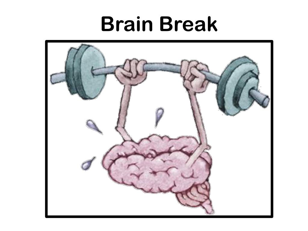
The spinal nerves are responsible for communication between the brain and the body. Motor neurons transmit signals from the brain to control muscle movement. Sensory areas carry signals from body parts to the brain to communicate information about heat, cold, pressure, pain, and limb position.
Nerve damage
Regardless of the cause of spinal cord injury, nerve fibers passing through the damaged area can also be affected. This leads to a deterioration in the functioning of the muscles and nerves located below the injury site. Damage to the thoracic or lumbar region can affect the functioning of the muscles of the trunk, legs, and internal organs (bladder and bowel control, sexual function). And neck injuries can affect hand movements and even the ability to breathe.
Common causes of spinal cord injury
The most common causes of spinal cord injury in the United States:
Traffic accidents. Crashes involving cars and motorcycles are the leading cause of spinal cord injury, over 40% annually.
Falls. Spinal cord injuries in the elderly (after 65 years) are usually associated with a fall. In general, statistics allocate ¼ of all cases to this reason.
Acts of violence. 15% of spinal cord injuries are caused by violence (including gunshot and stab wounds). Data from the National Institute of Neurological Disorders and Stroke.
Sports injuries. Professional sports carry many dangers, as well as active recreation, for example, diving in shallow water. 8% of back injuries fall under this article.
Alcohol. Every fourth injury is related to alcohol in one way or another.
Diseases. Cancer, arthritis, osteoporosis, and inflammation of the spinal cord can also cause damage to this organ.
Despite the fact that such injuries are usually caused by an accident, a number of risk factors have been identified, such as:
Gender. Statistically affected men are many times more. In the US, there are only 20% of women with similar and injuries.
Age. As a rule, injuries are received at the most active age - from 16 to 30 years. Road accidents remain the leading cause of injury at this age.
Love for risk and extreme. Which is logical, but the main thing is that in the first place, athletes and amateurs get injured when safety precautions are violated.
Diseases of bones and joints. In the case of chronic arthritis or osteoporosis, even a small back injury can be fatal to the patient.
After a spinal cord injury, patients face a large number of unpleasant consequences that can radically change their lives. When receiving such a serious injury, a team of specialists comes to the aid of the patient, including neurosurgeons, neurologists and doctors of the rehabilitation center.
Specialists of the Rehabilitation Center will offer a number of methods to control vital processes (bladder and intestines). A special diet will be developed to improve organ functions, which will help to avoid future kidney stones, urinary tract and kidney infections, obesity, diabetes, etc. Under the supervision of experienced physiotherapists, a program of physical exercises will be developed to improve the patient's muscle tone. You will receive detailed advice on skin care to avoid pressure sores, maintaining the functioning of the cardiovascular and respiratory systems. Specialists in the field of urology and infertility treatment can also be involved if necessary. Doctors will teach you how to deal with pain and depression. We are able to offer an integrated approach for the complete stabilization of the patient's condition.
Medical research:
Radiography. This is where the study should start. The pictures give a general picture of the situation, allow assessing the deformation of the spine, detecting fractures, dislocations of the bodies and processes of the vertebrae, and clarifying the level of damage.
Computed tomography (CT). CT scan gives more detailed information about the damaged area. When scanning, the doctor receives a series of cross-sectional images and provides a detailed study of the walls of the spinal canal, its membranes and nerve roots.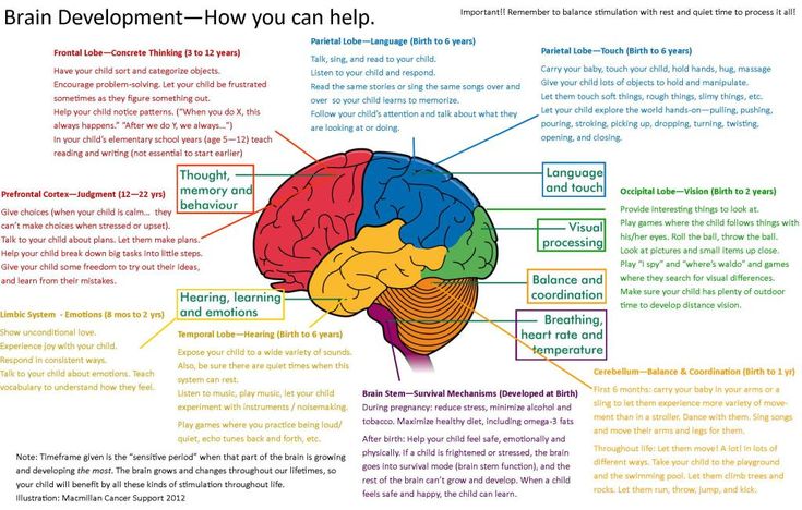
Magnetic resonance imaging (MRI). MRI makes it possible to obtain an image of the spinal cord throughout in different projections. And it will be very useful in identifying herniated discs, blood clots and other masses that can compress the spinal cord.
A few days after the injury, when the swelling has subsided, the doctor may do a neurological examination to determine the severity of the injury. It includes a test of muscle strength and sensory sensitivity.
Unfortunately, spinal cord injury cannot be completely cured. But ongoing research is providing physicians with more and more new tools and techniques to treat patients that can help regenerate nerve cells and improve nerve function. At the same time, we should not forget about the work that is being done in the field of maintaining an active life of patients after an injury, expanding opportunities and improving the quality of life of people with disabilities.
Ambulance service
Prompt first aid is critical to minimizing the consequences of any head or neck injury.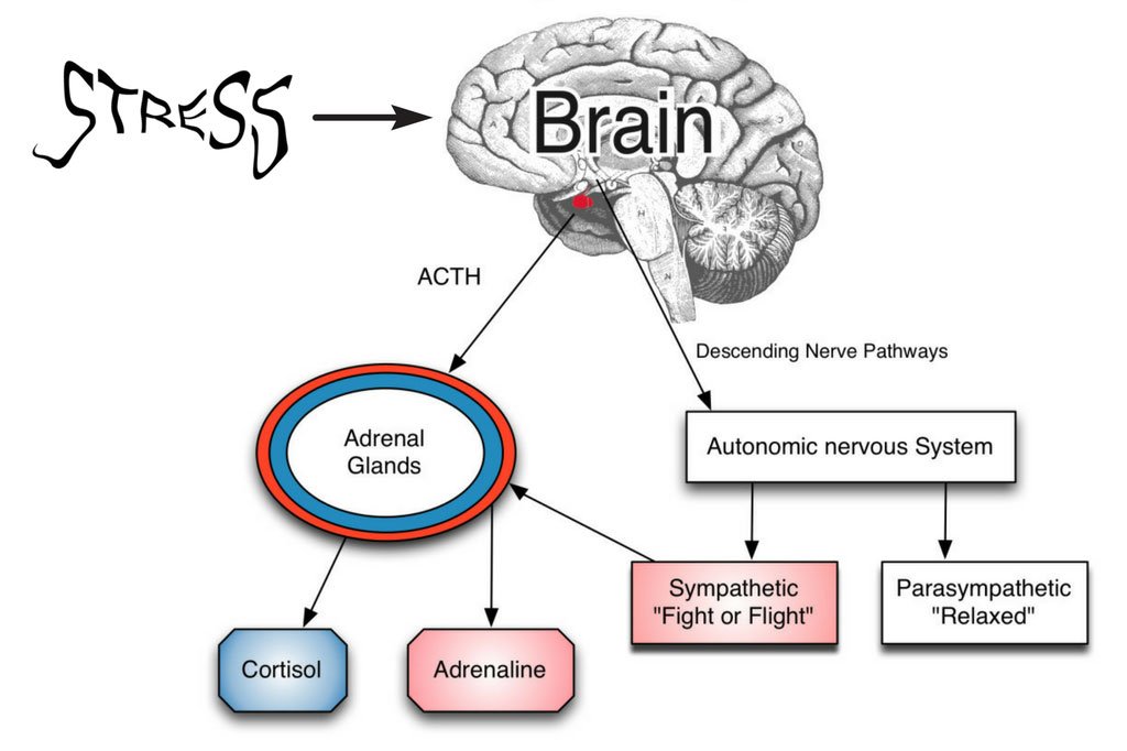 Similarly, spinal cord injury treatment often begins at the scene.
The emergency medical team on arrival should immobilize the spine as gently and quickly as possible using a rigid cervical collar and a special stretcher to transport the casualty to the hospital.
When a spinal cord injury occurs, the patient is taken to the intensive care unit. The patient can also be taken to a regional spinal injury center where a team of neurosurgeons, orthopedic surgeons, psychologists, nurses, therapists and social workers is always on duty.
Medicines. Methylprednisolone (Medrol) is used for acute spinal cord injury. When treated with "Methylprednisolone" within the first eight hours after injury, there is a chance to get a moderate improvement in the patient's condition. This drug reduces damage to nerve cells and relieves inflammation of the tissues around the site of injury. However, it is not a cure for spinal cord injury itself.
Immobilization. Stabilization of the injured spine during transportation is extremely important. To do this, the brigade has in its arsenal special devices for keeping the spine and neck stationary.
Surgical intervention. Often, doctors are forced to resort to operations to remove fragments of bones, foreign objects, herniated discs, or fix a fractured vertebrae. Surgery may also be needed to stabilize the spine to prevent pain or bone deformity in the future.
Period of hospitalization
Once the patient has been stabilized and treated as a matter of priority, the staff begins to work to prevent complications and associated problems. This may be a deterioration in the patient's physical condition, muscle contracture, bedsores, disruption of the intestines and bladder, respiratory infections and blood clots.
The length of hospital stay depends on the severity of the injury and the rate of recovery. After discharge, the patient is sent to the rehabilitation department.
Rehabilitation. Work with the patient can begin in the early stages of recovery. A team of specialists may include physical therapists, occupational therapists, specially trained nurses, a psychologist, a social worker, a nutritionist and a supervising physician.
In the early stages of rehabilitation, physicians typically work to preserve and strengthen muscle function by engaging fine motor skills and teaching adaptive behaviors in daily activities. Patients receive advice on the consequences of injuries and the prevention of complications. You will be given recommendations on how you can improve the quality of life in the current conditions. Patients are taught new skills, including the use of special equipment and technologies that make it possible not to depend on outside help. Having mastered them, you can find a possibly new hobby, participate in social and sports activities, return to school or work.
Medical treatment. The patient may be prescribed medication to control the effects of spinal cord injury. These include medicines to control pain and muscle spasms, as well as medicines to improve bladder, bowel and sexual function control.


.png)